Everyone knows the sensation of feeling the own heart beating – be it because you speak for the first time to the love of your life, or you give a speech in front of a big crowd, or sprint to reach a bus and are out of breath. Although investigation of how the brain controls and processes such heart activity has become a prominent topic in recent years, the question remained unresolved which brain structures are responsible for the sensation of the “beating heart”. Particularly the cardiac field artifact (CFA) that originates from the myocardial muscle contaminates electroencephalography (EEG) recordings and handicaps electrophysiological research in this direction, masking the true heart beat-related brain responses.
Markus Kern and his colleagues at the Faculty of Biology, the Epilepsy Center at the University Medical Center, and the Bernstein Center Freiburg now used a new approach to investigate heart cycle-related effects in the human brain. In electrocorticographic (ECoG) recordings, electrodes are placed directly on the surface of the human brain. The scientists used recordings from epilepsy patients during natural behavior, i.e., the patients were neither instructed to attempt to consciously perceive their heartbeats nor to ignore the perception. This allowed collecting a large number of trials without additional burden on the patients.
The team of researchers observed different types of heart cycle-related ECoG responses and compared them to their counterparts in EEG. And in fact, they found for the first time a focally localized heart beat-related brain response over the primary sensory cortex, observable clearly and consistently over all subjects investigated.
“This means that even if we don’t consciously perceive our heartbeat, the major somatosensory area of our brains still permanently responds to our heart beats”, explains Markus Kern. The scientist hopes that research on people consciously perceiving their heartbeat will help to identify entire neural networks involved in the processing of cardiac related signals. He is confident that their method of recording directly from the brain surface is the way to go: “ECoG with its high signal to noise ratio (SNR), its excellent temporal and spatial resolution seems to be an optimal candidate for further research in this direction. Beyond that, taking into account heart-beat related effects can increase the signal quality of ECoG even further and help both scientific studies and new biomedical applications based on the ECoG, such as brain-machine interfaces (BMIs).”
Image caption
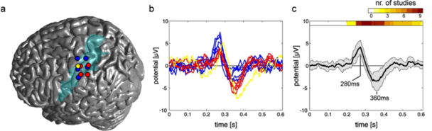
Location and time course of the heartbeat evoked potential HEP. (a) Colored circles (colors indicate individual subjects) correspond to electrode contacts over the somatosensory cortex (blue area) that recorded HEPs. (b) Corresponding time course of the individual HEPs and grand average with standard deviation (c). At the top of the graph are HEP latencies of previous EEG studies color encoded.
Original article
Markus Kern, Ad Aertsen, Andreas Schulze-Bonhage and Tonio Ball (2013) Heart cycle-related effects on event-related potentials, spectral power changes, and connectivity patterns in the human ECoG. Neuroimage 81, 178-190